Ask why a pink Alocasia turned green and you'll get two confident answers. One says give it more light. The other says light has nothing to do with it. Both of them are describing something real. They're describing different plants, and one word is doing the work of four.
One Word, Four Causes
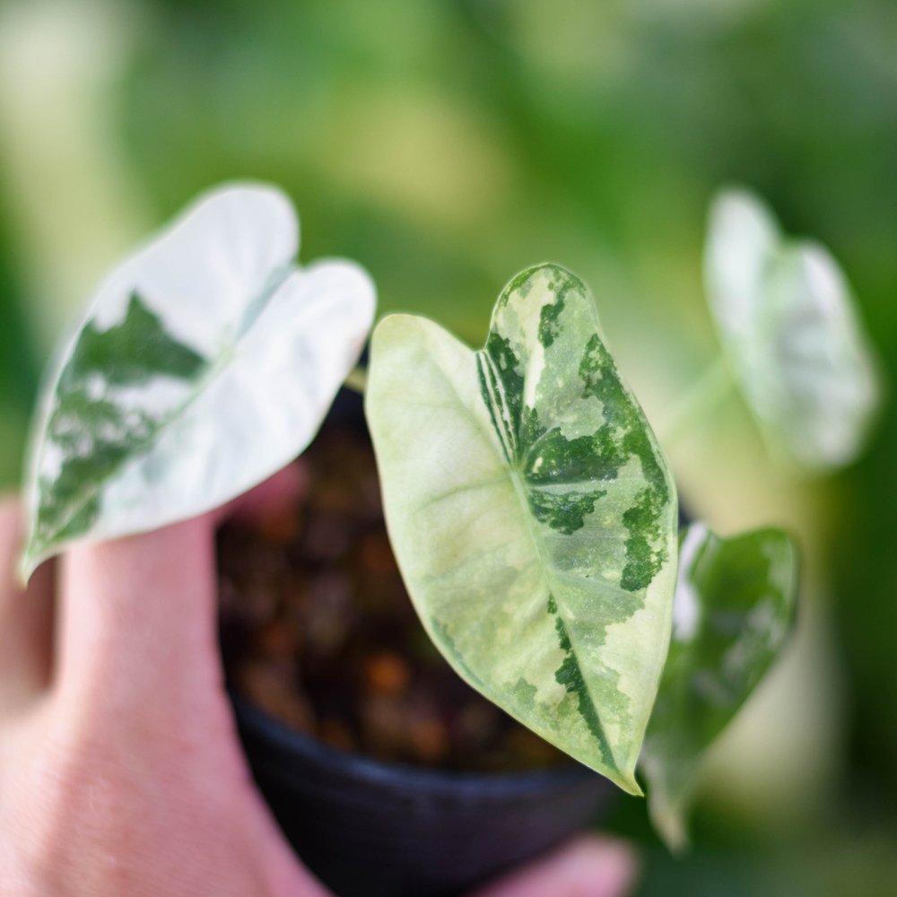
Four unrelated things make an Alocasia leaf something other than plain green. They come from different causes, and only one answers to light at all. Just one of them is variegation in the strict sense, and it isn't the one most people are trying to manage.
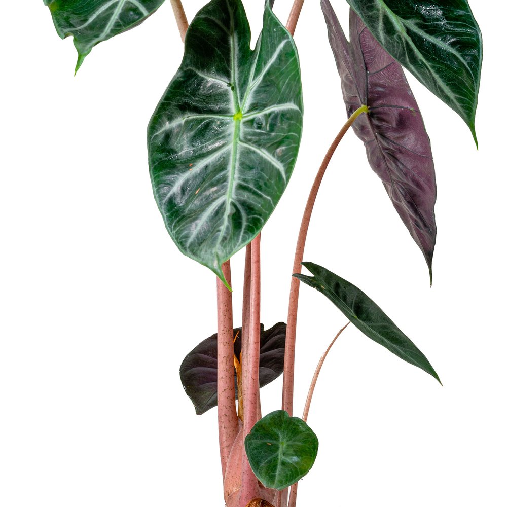
Pigment. The plant manufactures a red-to-purple compound and stores it in its cells. It can make more, and it can stop. This one responds to light.
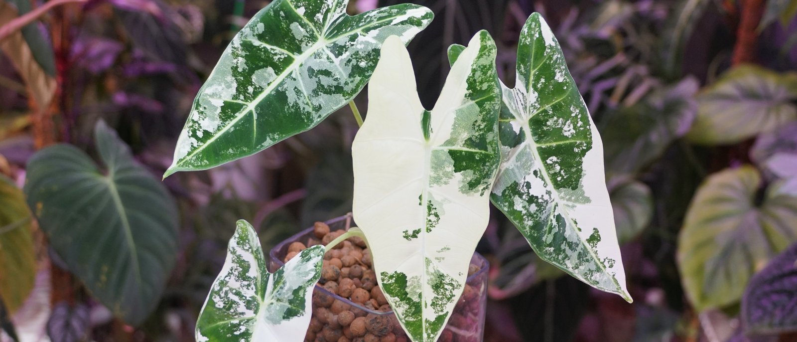
Architecture. The growing tip is built from stacked layers of cells, and in some plants those layers carry different genetics. Pale sectors come from a layer that can't build chlorophyll. This is the one that earns the word variegation, and light does nothing to it.
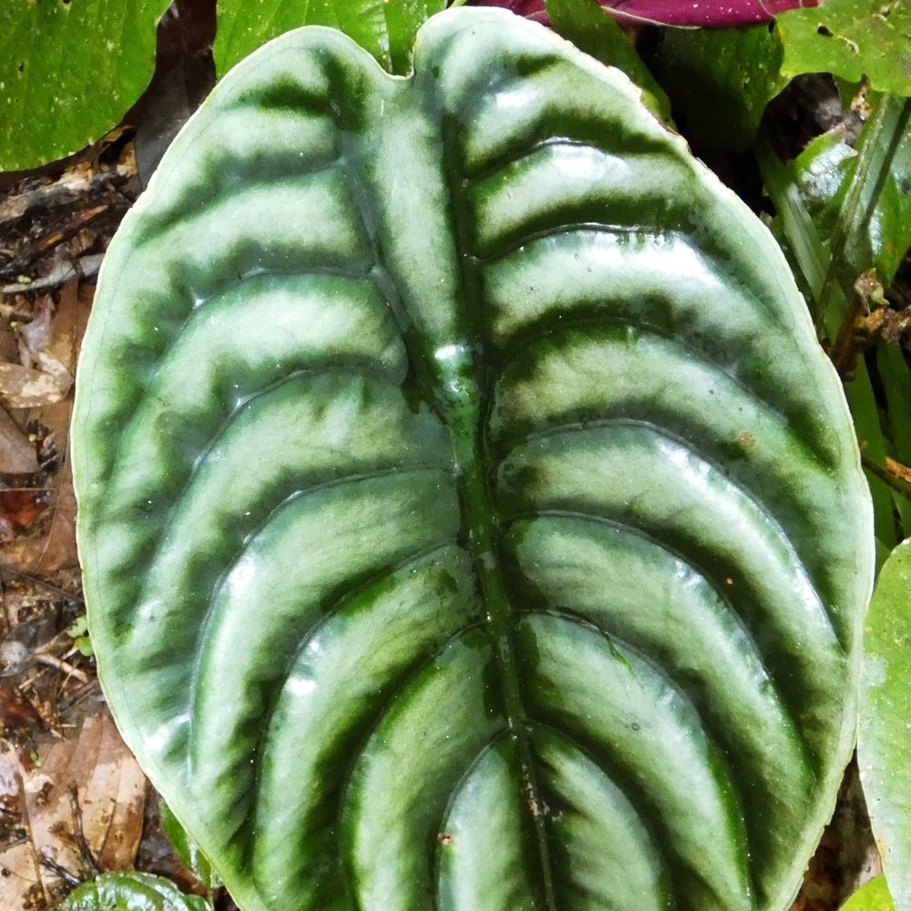
Structure. The silver of a Dragon Scale and the copper of an A. cuprea aren't pigment in the ordinary sense. They're an optical effect of how the tissue is built. The mechanism in aroids is unidentified, and not for want of looking.
Infection. Pale mottling is sometimes a virus.
1 · Pigment: What Anthocyanin Actually Does
Anthocyanins are the compounds behind red, purple and pink in plant tissue. They sit in the vacuole, the storage compartment inside a cell, which often puts them in front of the chloroplasts doing the photosynthesis, acting like a filter. Not always, though. When Gould and colleagues surveyed a thousand leaves of one naturally variable species, they found anthocyanins in almost every tissue combination the leaf offered, most commonly inside the photosynthetic cells themselves (Gould et al., 2000).
Anthocyanin is manufactured. The plant builds it, at a cost, in response to conditions. Steyn and colleagues put it plainly in their 2002 review: accumulation requires light, and tends to show up when the plant is taking in more light than it can currently put to work.
Two more levers push it up, and they're the ones you can actually pull. Cold does it: in tomato, dropping growth temperature from 75 °F to 64 °F or 54 °F (24 °C to 18 °C or 12 °C) produced pronounced increases in anthocyanin specifically (Løvdal et al., 2010). Lean feeding does it too, though that tomato work is careful about which compounds respond to what. Nitrogen shortage raised flavonoids and caffeoyl derivatives, while the anthocyanin response tracked temperature. For nitrogen acting on anthocyanin directly, the cleaner evidence is Oh and colleagues (2014). They found that shortages of nitrogen and phosphate, and cold, all push anthocyanin up through the same light-sensing machinery in the leaf. Red light drove it harder than blue when the plants were cold. Løvdal's team also found these factors compound each other rather than simply adding up.
Notice what light, cold and lean feeding have in common. Every one is a mild stress signal. A plant pushed for color is a plant held slightly below its comfortable optimum. That's a trade, not a free win.
Does It Protect the Leaf, or Expose It?
This is where the internet contradicts itself, and it does so because the primary literature does too.
The case for protection is strong. Steyn's team proposed a unified photoprotective function: the pigment attenuates light, which reduces the mismatch between energy coming in and carbon the plant can fix, which lowers the risk of oxidative damage. Zhao and colleagues (2022) overexpressed an anthocyanin regulator in apple and concluded the pigment does shield the leaf from high light stress. Their paper opens by conceding the role "remains controversial," and their own results show why. Photoinhibition is the slowdown a leaf takes when it catches more light than it can put to work. Out in the field, where photosynthesis was running normally, the red leaves showed just as much of it as the green ones. The protection is real, and it's conditional.
One sub-argument is settled. Anthocyanins are good antioxidants in a test tube, and for years that was offered as the mechanism. Zheng and colleagues (2021) tested the two explanations against each other in Arabidopsis and concluded that light attenuation matters more than antioxidant activity. The antioxidant story isn't wrong so much as secondary.
Here's the sub-claim you'll actually hear most, and it needs the same treatment: that a pink leaf is sunscreen against ultraviolet light. Mostly it isn't, and the mislabel is worth fixing. In a leaf, ultraviolet screening is chiefly the work of colorless flavonoids, a different set of compounds from the anthocyanin, and a role pinned down with mutant plants that can't build them and turn ultraviolet-sensitive as a result (Agati et al., 2021). Anthocyanin's own contribution is to visible light. It absorbs across the blue, green and red bands on their way to the chloroplasts, which is real and useful, but it is not the ultraviolet shield the folk version promises. One caution against overcorrecting, from that same review: anthocyanin isn't "just a green pigment" either, a line repeated about as often as the sunscreen one. In the living leaf it takes up blue and red light too, and that visible-light filtering is exactly what the protection argument here rests on (Agati et al., 2021).
A filter costs you light you were going to use. Gould, Vogelmann, Han and Clearwater (2002) measured photosynthesis through red and green leaves of Quintinia serrata. Under the pigment layer the mesophyll, the leaf's inner photosynthetic tissue, took on the characteristics of shade-adapted tissue, and the red leaves showed a 23 percent reduction in carbon dioxide assimilation at light saturation.
That result keeps reappearing. Anthocyanin-accumulating tomato had thinner leaves and lower photosynthetic rates alongside its extra protection, which the authors named a trade-off between photoprotection and growth (Cerqueira et al., 2023). Purple basil tolerated strong light better than green basil but was "far less able to accommodate a large change in irradiance," and green basil was the better performer in shade (Tattini et al., 2014).
So anthocyanin isn't protective or harmful in the abstract. It's a filter with a running cost, paying under excess light and charging rent under low light. Most of the conflicting claims you'll find are two accurate observations made at different light levels.
One finding lines up unusually well with a shelf plant. That thousand-leaf survey found anthocyanin was most abundant in older leaves under canopy gaps, and the authors' conclusion is more skeptical than most summaries admit: the pigments "are associated with photosynthesis, but do not serve an auxiliary phytoprotective role. They may serve to protect shade-adapted chloroplasts from brief exposure to high intensity sunflecks" (Gould et al., 2000). A sunfleck is the patch of direct sun that swings across a forest floor when the canopy moves. Alocasias are understory plants, and a shade-built leaf handling short violent bursts of light is exactly their problem. It's still an inference from a New Zealand tree to a Bornean aroid.
2 · Architecture: Why Chimeras Don't Answer to Light
Now the mechanism that actually is variegation, and the reason so much advice misfires.
The growing tip of a plant is built in layers. In most dicots there are three: the outermost builds the skin of every leaf it produces, the next builds most of the interior, and a third builds the core. Monocots, which is what an Alocasia is, are usually simpler, but the principle holds. Those layers stay separate because of the direction their cells divide. When they carry different genetics you get a chimera: one plant, two cell lines, stacked.
If one layer can't build chlorophyll, the tissue it produces comes out cream or white. That's the cream sectoring on a variegated Frydek. The pattern isn't a pigment the plant is making. It's a map of which layer built which part of the leaf.
Tilney-Bassett's Plant Chimeras (1986) laid out the framework, and Lineberger and Druckenbrod (1985) demonstrated it on pinwheel-flowered African violets, where the striped petals come from the outer and inner layers carrying different genetics. Both are eudicots, so read them as the model rather than as evidence about your Alocasia. The nearest aroid case is a laboratory one: Li and colleagues (2004) transformed Caladium bicolor and got a plant that came out symmetrically half green and half red, apparently from a single division that put one transformed and one ordinary cell side by side at the growing point. Engineered rather than natural, but it shows the architecture is available to aroids.
Here's the consequence. Reversion happens when the arrangement of those layers changes at the growing tip and one cell line takes over. In the chimera literature that's a lineage event, and light doesn't figure as a lever on it. If a shoot is throwing all-green growth, more light won't bring the pattern back. What light does change is vigor, and how visible the contrast is. Worth having, but not the same as holding the pattern.
The pattern travels with the growing tip. Propagate from growth that still carries both cell lines. If a shoot has gone fully green, its growing tip has lost the other line and will keep producing green.
This is also part of why plants bought from tissue culture arrive unpredictable. Routes that rebuild a shoot from scratch can lose or reshuffle the layer arrangement, while routes carrying an existing growing tip forward are better at preserving it (George et al., 2008). The picture isn't uniform: Norris and colleagues (1983) found shoots rebuilt from scratch that kept the parent pattern anyway, apparently because the new buds were founded by more than one cell. Tissue culture is good at producing pathogen-free stock. It doesn't guarantee a pattern, or an identical plant.
3 · Structure: The Jewels, and an Honest Gap
The silver of A. baginda, the copper sheen of A. cuprea. Not chlorophyll loss, and it doesn't revert. It's how these species are built.
Structural color in plants is well described. Ordered layers in the tissue reflect particular wavelengths, and the result is typically brighter than pigment can manage (Vignolini et al., 2013). Two of the known structures turn up in shade plants people actually grow, and they disagree about whether the color does anything useful. In Begonia, Jacobs and colleagues (2016) worked on the modified chloroplasts called iridoplasts, whose membranes are stacked in a regular photonic arrangement, and measured a 5 to 10 percent improvement in how efficiently those leaves turned dim light into chemistry. In Selaginella, Thomas and colleagues (2010) traced blue iridescence to a layered structure in the cuticle and reported that their results "do not support the idea that iridescence in plants acts to enhance light capture of photosynthetically important wavelengths." The likeliest reading is that these aren't in conflict but are different structures, one inside the light-harvesting machinery and one on the leaf surface. That's an inference, not something either paper states.
Now the aroid part, which is the interesting bit. A 2024 survey of structurally colored leaves across land plants examined Araceae directly. Despite some indication of structural color being present, the authors report that "preliminary investigations have not revealed photonic helicoids" in the aroids they looked at (Lundquist et al., 2024). Helicoidal cell walls are the commonest mechanism for structural color in leaves, and that team raised the count of genera carrying them to at least 35, every one a fern or a monocot in the orchid family or three families of Poales. Their conclusion is blunt: "all other angiosperms apparently lack photonic helicoidal cell walls in their leaves."
So aroids have been looked at, the commonest mechanism isn't the answer, and whatever produces the sheen on a Dragon Scale hasn't been identified. That's better than an absence of research. It's a narrowed question. Which also means that when you read that velvet leaves are an adaptation for capturing light in deep shade, the claim is measured in Begonia, contradicted in Selaginella, and unresolved in every Alocasia anyone has grown. Lundquist's team are careful too, noting the advantage "remains unclear, particularly as they reflect high-energy blue light that might otherwise be used for photosynthesis."
Two cautions. Velvet and metallic aren't the same phenomenon: a Black Velvet and a copper cuprea are doing different optical things, and neither has been measured. And the near-black ground of A. reginula may not belong in this bucket at all. If that darkness turns out to be anthocyanin it belongs in the first category, and it would answer to light. Nobody has published either way.
4 · Infection: When the Pattern Is a Problem
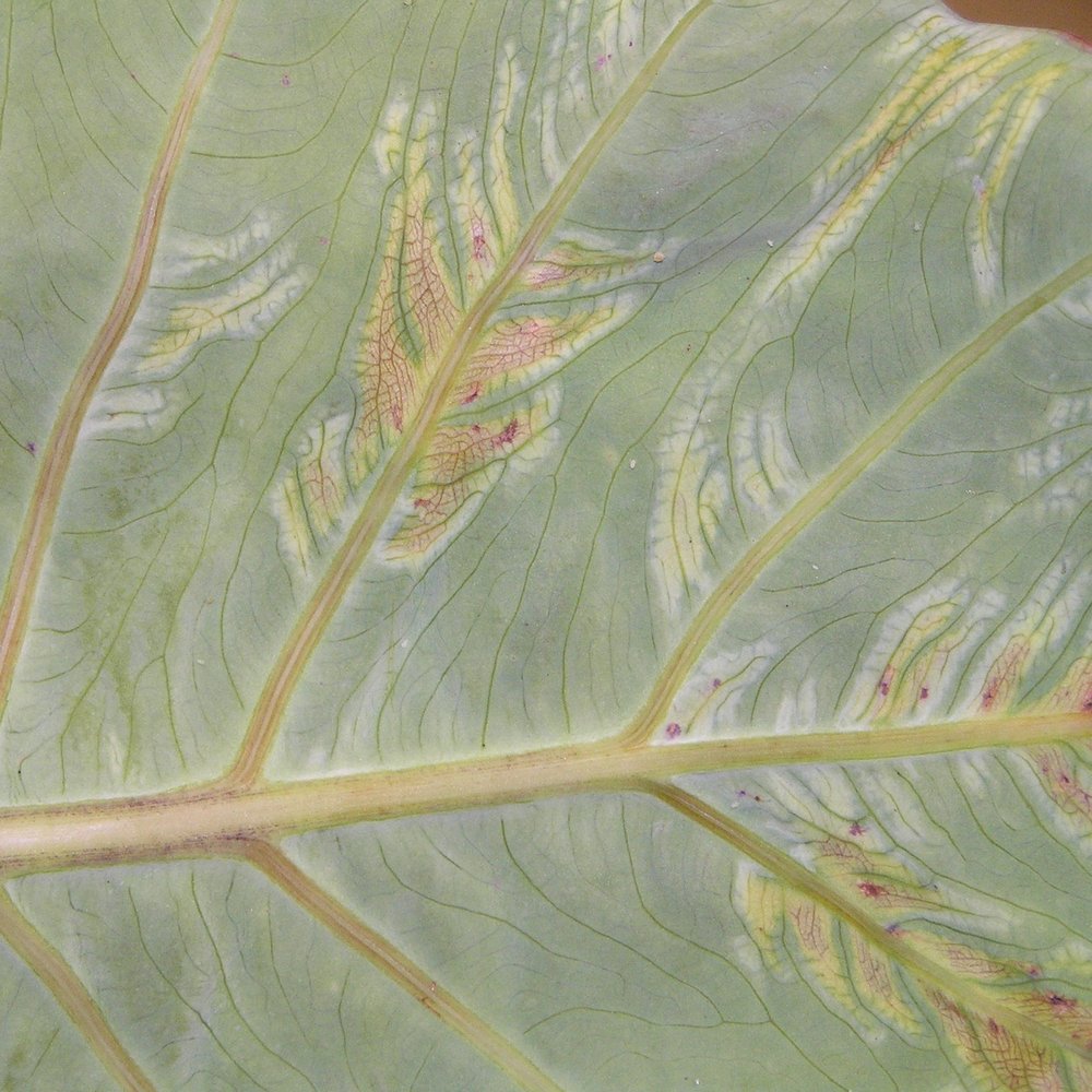
Dasheen mosaic virus is an aphid-transmitted virus that infects cultivated aroids worldwide. Alocasia is a documented host, and Nelson's extension datasheet (2008) files it under systemic mosaic. That distinction matters, because the chlorotic feathering, meaning pale yellow streaking, along leaf veins that people most often picture is listed for Colocasia and Xanthosoma, not for Alocasia. How the symptoms show varies with the host, the strain and the growing conditions. They can come and go, and greenhouse ornamentals sometimes show nothing at all.
Management, in Nelson's terms, rests on clean planting material and controlling the aphids that move the virus around. There's no treatment that clears an infected plant.
The practical tell here is weaker than I'd like, and I'd rather say so than hand you false confidence. Chimeral variegation follows sectors, with boundaries that respect the geometry of how the leaf was built. Viral mosaic tends to look diffuse and irregular. But Nelson notes that Philodendron vein clearing can resemble chlorotic feathering, and his recommended route to an actual answer is a laboratory test rather than an eye. The thing worth noticing is a new pale pattern on a plant that never had one.
Do Pink Leaves Resist Pests?
Short answer: nobody has tested this on an aroid, and the honest reading of the adjacent evidence is no.
The best evidence that leaf color deters herbivores is a signaling story rather than a chemical one. Cooney and colleagues (2012) showed that red-margined Pseudowintera colorata leaves contained more polygodial, a compound that discourages feeding, and took less damage in the wild. In feeding trials, caterpillars ate more of the green-margined tissue under normal light but showed no preference when the light was adjusted so they couldn't tell the colors apart.
Read that carefully. The defense was the polygodial. The red was the advertisement. That arrangement needs two things: a genuine chemical deterrent that tracks the color, and a herbivore that can see color and cares. Neither is established for a pink Alocasia. The genus's chemical defense is calcium oxalate, needle-shaped raphide crystals held in specialized cells throughout the plant and released when tissue is damaged (Mayo et al., 1997). They're there whatever color the leaf is.
One bad argument is worth retiring. The idea that houseplant pests can't see color is false: behavioral work on western flower thrips points to a three-receptor system covering ultraviolet, blue and green (Stukenberg et al., 2020).
Where an effect has been measured on pigmented ornamental foliage, it tracked leaf chemistry rather than appearance, and the result rewards careful reading. On a red-and-green Acalypha, spider mites developed faster and laid more eggs on the green form, which carried more chlorophyll, carotenoids, carbohydrate and soluble sugars. Phenol and anthocyanin content went the other way, correlating with longer male development and a longer life cycle (Safar & Mohamed, 2024). So the pigmented form was the poorer host, by nutrition rather than by camouflage.
For chimeras specifically the expectation runs the other way again. Less chlorophyll means less carbon coming in, and less in the bank to rebuild a leaf that gets chewed.
What This Means for Your Plant
Start by working out which of the four you're looking at, because the right move is different for each.
If your pink is pigment, brighter light will deepen it, and so will slightly cooler nights and a lighter hand with nitrogen. Understand what you're buying: a filter over the chloroplasts, with a measurable cost to growth, on a plant that evolved in the understory. Push the light too hard and the pigment won't make up the difference.
If your pattern is chimeral, light is the wrong lever entirely. Keep growth steady rather than fast, propagate from balanced growth, and prune back shoots that have gone fully green before they take over the plant.
If it's the silver or the copper, do nothing. It's not going anywhere, and nobody yet knows what it's for.
And if the pattern arrived on its own, on a plant that never had one, treat that as a question rather than a windfall.
And if a leaf comes out solid pink or solid pale, with no green at all, work out which of two things you're seeing, because they pull in opposite directions. On a variegated plant it's the mirror of green reversion: the colorless cell line has taken the growing tip and built a whole leaf with little or no chlorophyll. Striking, and costly. The other case is pigment, not architecture. A new leaf flushing deep uniform pink from anthocyanin is doing something ordinary, and it usually greens up as it matures and lays down chlorophyll. The tell is the tissue itself. Cream-to-white means chlorophyll is missing, the chimeral case; saturated pink-to-red sitting over a still-working green leaf is pigment on top, and it isn't a warning.
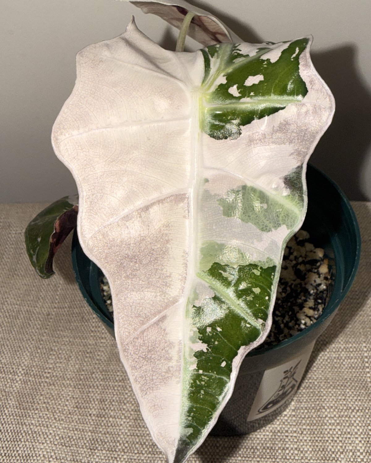
One more thing, and it's the least popular idea here. A leaf with no chlorophyll fixes no carbon. It isn't paying for itself. It's drawing on reserves the rest of the plant built. One such leaf is a curiosity. A plant producing nothing else is spending down its account. The right response is the opposite of the instinct: cut back to a node carrying both cell lines and let it push growth that can feed itself. You lose the spectacular leaf and keep the plant that can make more of them.
The Takeaway
The reason the advice out there conflicts is that half of it is about a pigment and half is about an architecture, and almost nobody says which. Pigment is something the plant makes and can stop making, and it answers to light, temperature and feeding. Architecture is an accident of which cells are building the plant, and it answers to none of those. The jewel sheen is a third thing, and its mechanism is still an open question in this family. A pattern that shows up uninvited is worth a second look. Sort out which one is on your shelf and the rest of the decisions make themselves.
Agati, G., Guidi, L., Landi, M., & Tattini, M. (2021). Anthocyanins in photoprotection: Knowing the actors in play to solve this complex ecophysiological issue. New Phytologist, 232(6), 2228–2235. https://doi.org/10.1111/nph.17648
Cerqueira, J. V. A., Zhu, F., Mendes, K., Nunes-Nesi, A., Martins, S. C. V., Benedito, V., Fernie, A. R., & Zsögön, A. (2023). Promoter replacement of ANT1 induces anthocyanin accumulation and triggers the shade avoidance response through developmental, physiological and metabolic reprogramming in tomato. Horticulture Research, 10(2), uhac254. https://doi.org/10.1093/hr/uhac254
Cooney, L. J., van Klink, J. W., Hughes, N. M., Perry, N. B., Schaefer, H. M., Menzies, I. J., & Gould, K. S. (2012). Red leaf margins indicate increased polygodial content and function as visual signals to reduce herbivory in Pseudowintera colorata. New Phytologist, 194(2), 488–497. https://doi.org/10.1111/j.1469-8137.2012.04063.x
George, E. F., Hall, M. A., & De Klerk, G.-J. (Eds.). (2008). Plant propagation by tissue culture: Vol. 1. The background (3rd ed.). Springer. https://doi.org/10.1007/978-1-4020-5005-3
Gould, K. S., Markham, K. R., Smith, R. H., & Goris, J. J. (2000). Functional role of anthocyanins in the leaves of Quintinia serrata A. Cunn. Journal of Experimental Botany, 51(347), 1107–1115. https://doi.org/10.1093/jexbot/51.347.1107
Gould, K. S., Vogelmann, T. C., Han, T., & Clearwater, M. J. (2002). Profiles of photosynthesis within red and green leaves of Quintinia serrata. Physiologia Plantarum, 116(1), 127–133. https://doi.org/10.1034/j.1399-3054.2002.1160116.x
Jacobs, M., Lopez-Garcia, M., Phrathep, O.-P., Lawson, T., Oulton, R., & Whitney, H. M. (2016). Photonic multilayer structure of Begonia chloroplasts enhances photosynthetic efficiency. Nature Plants, 2(11), 16162. https://doi.org/10.1038/nplants.2016.162
Li, S. J., Deng, X. M., Mao, H. Z., & Hong, Y. (2004). Enhanced anthocyanin synthesis in foliage plant Caladium bicolor. Plant Cell Reports, 23(10–11), 716–720. https://doi.org/10.1007/s00299-004-0871-2
Lineberger, R. D., & Druckenbrod, M. (1985). Chimeral nature of the pinwheel flowering African violets (Saintpaulia, Gesneriaceae). American Journal of Botany, 72(8), 1204–1212. https://doi.org/10.1002/j.1537-2197.1985.tb08373.x
Lundquist, C. R., Rudall, P. J., Sukri, R. S., Conejero, M., Smith, A., Lopez-Garcia, M., Vignolini, S., Metali, F., & Whitney, H. M. (2024). Living jewels: Iterative evolution of iridescent blue leaves from helicoidal cell walls. Annals of Botany, 134(1), 131–150. https://doi.org/10.1093/aob/mcae045
Løvdal, T., Olsen, K. M., Slimestad, R., Verheul, M., & Lillo, C. (2010). Synergetic effects of nitrogen depletion, temperature, and light on the content of phenolic compounds and gene expression in leaves of tomato. Phytochemistry, 71(5–6), 605–613. https://doi.org/10.1016/j.phytochem.2009.12.014
Mayo, S. J., Bogner, J., & Boyce, P. C. (1997). The genera of Araceae. Royal Botanic Gardens, Kew.
Nelson, S. C. (2008). Dasheen mosaic of edible and ornamental aroids (PD-44). University of Hawaiʻi College of Tropical Agriculture and Human Resources. https://www.ctahr.hawaii.edu/oc/freepubs/pdf/PD-44.pdf
Norris, R., Smith, R. H., & Vaughn, K. C. (1983). Plant chimeras used to establish de novo origin of shoots. Science, 220(4592), 75–76. https://doi.org/10.1126/science.220.4592.75
Oh, S., Warnasooriya, S. N., & Montgomery, B. L. (2014). Mesophyll-localized phytochromes gate stress- and light-inducible anthocyanin accumulation in Arabidopsis thaliana. Plant Signaling & Behavior, 9(1), e28013. https://doi.org/10.4161/psb.28013
Safar, S. H. M., & Mohamed, I. A. A. (2024). Interaction between biological aspects of Tetranychus urticae Koch (Acari: Tetranychidae) and some chemical composition in two colored Acalypha wilkesiana Müll. Arg. (Malpighiales: Euphorbiaceae) leaves. Persian Journal of Acarology, 13(3), 499–512. https://doi.org/10.22073/pja.v13i3.85113
Steyn, W. J., Wand, S. J. E., Holcroft, D. M., & Jacobs, G. (2002). Anthocyanins in vegetative tissues: A proposed unified function in photoprotection. New Phytologist, 155(3), 349–361. https://doi.org/10.1046/j.1469-8137.2002.00482.x
Stukenberg, N., Pietruska, M., Waldherr, A., & Meyhöfer, R. (2020). Wavelength-specific behavior of the western flower thrips (Frankliniella occidentalis): Evidence for a blue-green chromatic mechanism. Insects, 11(7), 423. https://doi.org/10.3390/insects11070423
Tattini, M., Landi, M., Brunetti, C., Giordano, C., Remorini, D., Gould, K. S., & Guidi, L. (2014). Epidermal coumaroyl anthocyanins protect sweet basil against excess light stress: Multiple consequences of light attenuation. Physiologia Plantarum, 152(3), 585–598. https://doi.org/10.1111/ppl.12201
Thomas, K. R., Kolle, M., Whitney, H. M., Glover, B. J., & Steiner, U. (2010). Function of blue iridescence in tropical understorey plants. Journal of the Royal Society Interface, 7(53), 1699–1707. https://doi.org/10.1098/rsif.2010.0201
Tilney-Bassett, R. A. E. (1986). Plant chimeras. Edward Arnold.
Vignolini, S., Moyroud, E., Glover, B. J., & Steiner, U. (2013). Analysing photonic structures in plants. Journal of the Royal Society Interface, 10(87), 20130394. https://doi.org/10.1098/rsif.2013.0394
Zhao, S., Blum, J. A., Ma, F., Wang, Y., Borejsza-Wysocka, E., Ma, F., Cheng, L., & Li, P. (2022). Anthocyanin accumulation provides protection against high light stress while reducing photosynthesis in apple leaves. International Journal of Molecular Sciences, 23(20), 12616. https://doi.org/10.3390/ijms232012616
Zheng, X.-T., Yu, Z.-C., Tang, J.-W., Cai, M.-L., Chen, Y.-L., Yang, C.-W., Chow, W. S., & Peng, C.-L. (2021). The major photoprotective role of anthocyanins in leaves of Arabidopsis thaliana under long-term high light treatment: Antioxidant or light attenuator? Photosynthesis Research, 149(1–2), 25–40. https://doi.org/10.1007/s11120-020-00761-8